Inspiring Future, Grand Challenge
Prof. Kim Tae-Hyung
Development of a Nanocomposite-Based Electrochemical Platform
Prof. Woo Choong-Wan
Uncovering the neural mechanisms of pain processing
Prof. Kim, Hyunggoo
AI-driven identification of instinctive behaviors in living organisms
Spring 2021 IPHC Seminar Series
행사/세미나 Spring 2021 IPHC Seminar Series
Professor Taehyung Kim's Research Team Selected for NRF Individual Basic Research Program
Professor Taehyung Kim's Research Team Selected for NRF Individual Basic Research Program - Professor Tae-Hyung Kim has been selected for the Mid-Career Researcher Support Program (Type 1 - Global Cooperation) with his research on the development of a 3D nanomaterial-based multi-layer platform technology, receiving approximately ₩300 million per year for five years. - Dr. Kyeong-Mo Koo, a research fellow, has been awarded the Sejong Science Fellowship (Domestic Track) for his research on the development of a platform technology for cellular rejuvenation monitoring, receiving approximately ₩130 million per year for five years. ▲(From left) Professor Tae-Hyung Kim, Dr. Kyeong-Mo Koo ◇ Professor Tae-Hyung Kim and Dr. Kyeong-Mo Koo Selected for NRF Mid-Career Researcher Support Program and Sejong Science Fellowship The research team of Professor Tae-Hyung Kim has been selected for the NRF Mid-Career Researcher Support Program (Type 1 - Global Cooperation Project) under the NRF Individual Basic Research Program. Additionally, Dr. Kyeong-Mo Koo, a research fellow from the same lab, has been chosen for the Sejong Science Fellowship (Domestic Track). ◇ Professor Tae-Hyung Kim Selected for the NRF Mid-Career Researcher Support Program – Development of a 3D Nanomaterial-Based Multi-Layer Platform for Trilineage Formation The Mid-Career Researcher Support Program is designed to enhance the fundamental research capabilities of mid-career researchers and foster them into leading scientists by supporting their innovative research endeavors. Professor Kim’s research focuses on ‘Development of a three-dimensional nanomaterial multi-layer platform for high-efficiency autonomous generation of trilineage structures in vitro’. His project aims to control the differentiation of induced pluripotent stem cells (iPSCs) autonomously, facilitating the automatic formation and interaction of the three germ layers in a 3D environment. Expressing his gratitude, Professor Kim stated, “I am honored to be conducting this project among many outstanding researchers. I would like to extend my sincere appreciation to Sungkyunkwan University and the Industry-Academia Cooperation Foundation for their tremendous support during the proposal preparation process.” ▲[Figure 1] Development of a three-dimensional nanomaterial multi-layer platform for high-efficiency autonomous generation of trilineage structures in vitro ▲[Figure 2] Development of a Label-Free and Non-Destructive Multiplexing Electrochemical Platform for Mitochondrial Function Evaluation in Cellular Rejuvenation Monitoring
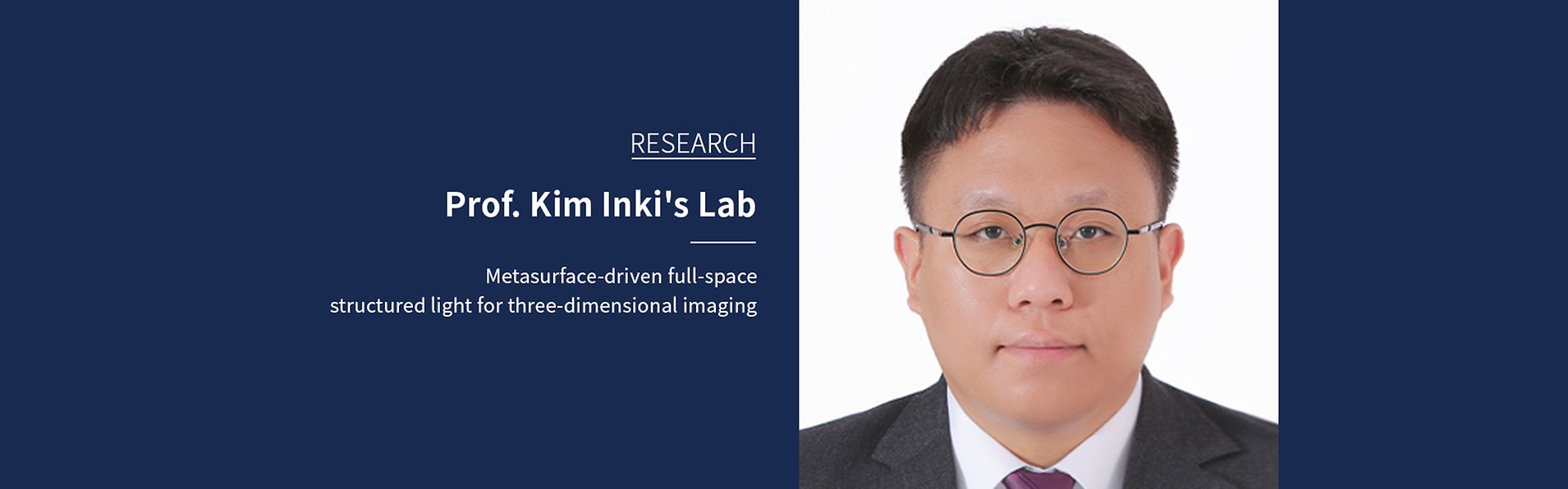
IPHC Welcomes New Assistant Professor
IPHC is pleased to introduce new faculty member, Professor Park Hangue, in the fall of 2022. After completing his bachelor's degree in electrical and computer engineering at Seoul National University, Professor Park Hangue gained experience in semiconductor integrated circuits during his master's and Samsung Electronics tenure and conducted research on capsule endoscopy. Later, he received a Ph.D. from the Georgia Institute of Technology for research on improving cat gait using closed neural stimulation. After that, as an assistant professor at Texas A&M University for 5 years, he conducted interesting studies on neural signal control and neural prosthetic system design using electrical stimulation. The main purpose of the study is to supplement human incomplete motor skills and rehabilitate after trauma, and has recently expanded its scope of use to enhance human and improve motor skills. The focus of the research is on designing semiconductor integrated circuit-based biomimicry to efficiently communicate with the nervous system and have minimal power consumption & size, and its effectiveness is thoroughly verified through human and animal experiments. Welcome to IPHC!
IPHC is pleased to introduce new faculty member, Professor Jung Seung Lee, in the spring of 2021. Dr. Jung Seung Lee grew up in Seoul, Korea and received his BA and PhD in the Department of Biotechnology at Yonsei University. Dr. Lee’s previous work focused on the development of novel functional biomaterials for successful stem cell engineering and tissue regeneration. For his post-doctoral research, he transitioned to the fields of translational engineering in the laboratory of Professor Robert Langer and Giovanni Traverso at Massachusetts Institute of Technology (MIT) where he developed a series of novel technologies for smart sensor and delivery platform for disease treatment. His current research interest is focused on developing the novel biomaterials for immune modulation and tissue reconstruction. Additionally, Dr. Jung Seung Lee continues his efforts towards the development of next generation of stem cell therapeutics. Welcome to IPHC!
Prof. Minah Suh gives a lecture in SKKU ExCampus Season 2
[This video provides subtitles in English and Chinese] Professor Minah Suh participated in SKKU ExCampus Season 2 hosted by the Student Success Center and gave a lecture under the title of "Another Universe! Looking at the Brain and Understanding the Brain.". More information can be found in SKKU News post (link).
Development of a Nanocomposite-Based Electrochemical Platform for Real-Time Mitochondrial Activity Analysis During Direc
Development of a Nanocomposite-Based Electrochemical Platform for Real-Time Mitochondrial Activity Analysis During Direct Reprogramming into Cardiomyocytes ▲(From left) Professor Tae-Hyung Kim, Dr. Kyeong-Mo Koo (Postdoctoral Fellow), Professor Yoonhee Jin (Yonsei University College of Medicine), and Seungju Seo (Ph.D. Candidate) - This study provides mechanistic insights into metabolic regulation during cellular reprogramming and holds promise for advancing cardiac regenerative therapeutics. - A joint research team led by Professor Tae-Hyung Kim at Sungkyunkwan University (SKKU) and Professor Yoonhee Jin at Yonsei University College of Medicine has developed a cutting-edge electrochemical platform capable of monitoring mitochondrial metabolic activity in real time during the direct reprogramming of cells into cardiomyocyte-like cells (CiCMs). The newly developed platform—a Poly-L-lysine/Matrigel double-layer coated gold nanostructured composite (PMGN)—enables quantitative, nondestructive analysis of mitochondrial function within 30 seconds, offering a powerful tool for tracking metabolic dynamics during cell fate conversion. Cardiovascular diseases remain the leading cause of death globally, and existing treatments largely focus on symptom management rather than restoring damaged heart tissue. While direct reprogramming has emerged as a promising regenerative approach, conventional methods for analyzing cellular function often require fixation or destruction of cells, involve complex procedures, and take hours to complete—limiting their utility in clinical settings. This new platform addresses these challenges by providing a rapid, label-free, and physiologically relevant method for assessing metabolic shifts during reprogramming, potentially paving the way for the development of novel regenerative therapies for cardiac repair. ▲[Figure 1] Schematic Overview of the Nanocomposite-Based Electrochemical Platform for Real-Time Monitoring of Mitochondrial Activity During Direct Cardiac Reprogramming - To address the limitations of existing analytical methods, the research team engineered a novel functional platform by applying a dual coating of poly-L-lysine and Matrigel onto gold nanostructured electrodes. This design significantly enhances both cell adhesion stability and electrochemical signal sensitivity, enabling reliable and sensitive metabolic measurements. The PMGN is capable of detecting the metabolic shift from glycolysis to oxidative phosphorylation (OXPHOS)—a hallmark of cardiomyocyte reprogramming—within approximately 30 seconds, in real time and without damaging the cells. Notably, the same cells can be repeatedly measured over time, offering a marked improvement in both efficiency and precision compared to conventional assays. Using this system, the team successfully tracked functional metabolic changes in reprogrammed cardiomyocyte-like cells over a long-term culture period of up to 29 days. Furthermore, the platform enabled real-time, label-free, and nondestructive assessment of cardiotoxic responses to four different compounds: doxorubicin, remdesivir, rosiglitazone (known cardiotoxic agents), and liraglutide (a non-toxic control). This achievement demonstrates the system’s potential not only for monitoring cell fate conversion but also for high-throughput cardiotoxicity screening in drug development. - This research presents broad applicability as a cardiotoxicity prediction platform for cardiovascular drug development, as well as for functional evaluation and quality control of cell-based therapeutics in regenerative medicine. It is expected to serve as a practical turning point in advancing cell therapy and drug development by providing a robust and scalable tool for assessing therapeutic efficacy and safety. - Keywords: Cell Adhesion Polymers, Gold Nanostructure, Electrochemical Method, Direct Cardiac Reprogramming, Mitochondrial Metabolism, Cardiotoxicity Assessment - Professor Kim remarked, “The PMGN platform, based on electrochemical mitochondrial activity analysis, enables quantitative assessment of cellular function without causing damage. It is expected to be widely applicable not only for quality control of cardiac regenerative cell therapies, but also for the screening of cardiotoxic drugs and evaluation of their clinical relevance.”He added, “I would like to express my sincere gratitude to SKKU and the Office of Research & Business Foundation for their generous support throughout this research.” - This research was supported by grants from the Ministry of Science and ICT and the Ministry of Health and Welfare of Korea through the National Research Foundation (NRF), the Regenerative Medicine Technology Development Program, the Korea ARPA-H, and the Korea Health Industry Development Institute (KHIDI). The findings were published on May 30, 2025, in Advanced Composites and Hybrid Materials (Impact Factor: 23.2), a leading international journal in the field of Materials Science and Composites (top 1.4% in the category). - Title: In situ monitoring of mitochondrial redox dynamics during cardiac reprogramming using a poly-l-lysine/Matrigel-coated gold nanostructured composite platform
New Study Reveals How the Brain Integrates Pain Prediction and Stimuli
New Study Reveals How the Brain Integrates Pain Prediction and Stimuli - Uncovering the neural mechanisms of pain processing using functional Magnetic Resonance Imaging (fMRI) - A study led by Associate Director WOO Choong-Wan of the Center for Neuroscience Imaging Research (CNIR) within the Institute for Basic Science (IBS), along with Michael YOO Seng Bum, Assistant Professor of Biomedical Engineering at Sungkyunkwan University, has uncovered new insights into how the brain processes and integrates pain information. Their research goes beyond identifying brain areas that respond to pain, revealing the mechanisms behind the brain's integration of pain-related information. Using functional magnetic resonance imaging (fMRI), they formalized how the brain combines pain expectations with the actual intensity of painful stimuli. Pain is a complex experience influenced not just by the intensity of a painful stimulus but also by the individual’s expectations. For instance, the pain one expects to feel can alter the perception of the actual pain experienced. While previous research has mapped out which brain regions handle these separate factors that contribute to our pain experience, this new study tackles the question of how these different factors come together to create a unified sensation of pain. KIM Jungwoo, the first author of the study, stated, “It’s not just about knowing which parts of the brain are important; ultimately, understanding how pain arises is key to figuring out how to eliminate unnecessary pain.” The researchers used fMRI to observe brain activity in participants exposed to varying levels of pain stimuli, while also manipulating their expectations about the level of pain they would feel. To fully understand how pain is processed in the brain, they separated the process into two stages: preservation (how the brain maintains information about pain expectations and stimulus intensity) and integration (how these elements combine to form a cohesive pain experience). They examined these processes across different levels of the brain’s cortical hierarchy*, expecting lower-level brain networks to preserve information without integrating it, and higher-level networks to preserve and integrate both. * Cortical Hierarchy: The brain processes information in a stepwise manner, with lower-level networks (like the sensory and motor networks) handling basic sensory input, and higher-level networks (such as the limbic system and default mode network) integrating more complex information. This study used this framework to understand how the brain processes and integrates pain information at different levels. Contrary to the researchers’ initial hypothesis, the results showed that all networks, regardless of level, preserved both types of information—pain expectations and stimulus intensity. However, only higher-level networks were able to integrate this information by simply adding the preserved expectation and stimulus information together. This suggests that while the entire brain stores pain information, only specific areas are responsible for integrating different pain-related signals into the experience of pain. This study represents a significant collaboration between two fields of neuroscience. Dr. Yoo, an expert in decision-making and electrophysiology, and Dr. Woo, a pain researcher specializing in fMRI, combined their expertise to explore how pain information is processed across the whole brain. Their innovative approach sheds light on the brain’s mechanisms for processing pain, providing valuable insights that could lead to new approaches to treating chronic pain. Michael YOO Seng Bum, the co-lead author said “It was a meaningful collaborative study that combined the strengths of each principal investigator to advance beyond merely reporting the activation of specific regions, allowing us to investigate principles of how information is integrated across the brain.” WOO Choong-Wan, another co-lead author, described the research as “an innovative study using geometric information encoded in brain activation patterns to reveal the integration mechanism of distinct types of pain information,” adding that “this discovery would not have been possible without a collaboration.” Figure 1. Hypothesis on the preservation and integration of pain information The left figure represents a three-dimensional space formed by fMRI voxels (volumetric activity unit captured by fMRI) within a network. The green two-dimensional plane (expectation subspace) represents the subspace preserving information about pain expectations, while the orange two-dimensional plane (stimulus subspace) represents the subspace preserving information about the stimulus intensity. The temporal activities of a network were projected onto these subspaces, and based on this information, the study examined whether each network preserved or integrated the two types of pain information. The initial hypothesis proposed that in lower-level networks (blue box), only one type of pain information would be well-preserved, leading to poor reconstruction of participants’ pain reports (indicating a lack of integration). In contrast, higher-level networks (red box) were hypothesized to preserve and integrate both types of pain information. Figure 2. Subspace patterns and comparison of reconstructed vs. actual pain reports Results are shown for the visual network (lower-level network) and the limbic system (higher-level network). The first row displays results from the visual network, while the second row shows results from the limbic system. From left to right, the columns represent brain patterns over time in the expectation subspace, brain patterns over time in the stimulus subspace, and a comparison between reconstructed pain ratings and actual pain ratings. The subspaces shown in the first and second columns preserved information related to each pain expectation and stimulus intensity. In the third column, the visual network primarily reconstructs differences in pain expectations, distinguished by different colors (red, gray, blue). In contrast, the limbic system successfully reconstructs both pain expectations and stimulus intensity information. Notes for editors - References Jungwoo Kim, Suhwan Gim, Seng Bum Michael Yoo, Choong-Wan Woo, A Computational Mechanism of Cue-Stimulus Integration for Pain in the Brain / Science Advances (2024) - Media Contact For further information or to request media assistance, please contact Choong-Wan Woo at the Center for Neuroscience Imaging Research, Institute for Basic Science (IBS) or Seng Bum Michael Yoo (CNIR) or William I. Suh at the IBS Public Relations Team (willisuh@ibs.re.kr). - About the Institute for Basic Science (IBS) IBS was founded in 2011 by the government of the Republic of Korea with the sole purpose of driving forward the development of basic science in South Korea. IBS has 7 research institutes and 31 research centers as of June 2024. There are eight physics, three mathematics, five chemistry, seven life science, two earth science, and six interdisciplinary research centers. Source : IBS Research News Link
HyungGoo Kim’s Joint Research Team Unveils Instinctive Behaviors in Living Organisms Using Artificial Intelligence
Professor HyungGoo Kim's Team from the Global Biomedical Engineering Department Uncovers Principles of Instinctive Behaviors Using Artificial Intelligence Revealing the Mechanisms of "Hunger" and "Appetite" in Hypothalamic Neurons with AI-Based Models Researchers led by Professor HyungGoo Kim from Sungkyunkwan University’s Global Biomedical Engineering Department (co-first author: master’s student Jong Won Yun) and Professor Hyung Jin Choi from Seoul National University (co-first authors: Ph.D. student Kyu Sik Kim, Dr. Young Hee Lee, and Ph.D. candidate Yu-Been Kim) have presented a novel method for understanding instinctive psychological states in humans through the application of artificial intelligence (AI). Despite advancements in neuroscience enabling the observation of diverse animal behaviors, the connection between neural signals and instinctive psychological states remains insufficiently understood. Although prior studies have associated specific hypothalamic neurons with instinctive behaviors, the precise roles and mechanisms of these neurons were unclear. This collaborative research provides the first quantitative analysis of hypothalamic neural functions using AI, thereby clarifying the relationship between instinctive psychological states and behaviors. The team combined a novel homeostatic theory with AI-based neural models to reveal that Agouti-related peptide (AgRP) neurons in the hypothalamus represent "hunger," while leptin receptor (LH LepR) neurons represent "appetite." Experimental observations of hypothalamic neural activity patterns were meticulously analyzed, experimentally demonstrating how hunger and appetite are encoded through the activity patterns of specific neural populations. Professor Kim utilized a computational modeling approach, initially developed to distinguish the roles of dopamine, to devise a methodology for differentiating neural activations. This method successfully expressed Professor Choi’s novel homeostatic theory in mathematical terms. Fusing Traditional Homeostatic Theories with Cutting-Edge Neuroscientific Discoveries Professor Kim stated, “This study is the first to quantitatively analyze the activities of complex neural circuits by integrating AI with neuroscience. It marks a pivotal step toward numerically understanding instinctive behaviors in living organisms. Particularly, our elucidation of how hypothalamic neural activity regulates basic instincts like hunger and appetite holds significant implications.” Professor Choi added, “Neural responses observed in the hypothalamus during studies on feeding behavior were difficult to explain using existing theories. AgRP neurons were activated to promote feeding but decreased their activity upon food presentation. Conversely, LH LepR neurons not only promoted feeding upon activation but also increased activity when food was provided. To understand these paradoxical findings, we employed AI models, which led to the establishment of a new homeostatic theory.” Modeling Process Utilizing Artificial Intelligence The study provides crucial insights into how the brain regulates survival-essential behaviors such as feeding. It is anticipated to pave the way for new strategies in treating eating disorders, obesity, and appetite-related conditions. By integrating AI into neuroscience, the research opens possibilities for quantifying and understanding human instinctive behaviors, offering a versatile approach to investigating other instinctive behaviors and psychological states. Key Experimental Results The study includes computer modeling to demonstrate how neural activity correlates with specific psychological elements. (Gray line: neural signals; red line: optimal hunger model; green line: optimal appetite model; blue and orange lines: control models). The joint research by Professor Hyunggoo Kim and Professor Hyung Jin Choi was published in Science Advances on November 6. Paper Title: A Normative Framework Dissociates Need and Motivation in Hypothalamic Neurons Journal: Science Advances DOI: https://doi.org/10.1126/sciadv.ado1820
Prof. Kim Inki's Lab published in Nature communications
Prof. Kim Inki's lab published 'Metasurface-driven full-space structured light for three-dimensional imaging' in Nature communications on October, 2022. Further details can be obtained from the following link. Kim, Gyeongtae, et al. "Metasurface-driven full-space structured light for three-dimensional imaging." Nature communications 13.1 (2022): 1-10.
Prof. Jang-Yeon Park's Lab published in Science
Prof. Jang-Yeon Park published 'In vivo direct imaging of neuronal activity at high temporospatial resolution' in Science on October, 2022. Further details can be obtained from the following link. Toi, Phan Tan, et al. "In vivo direct imaging of neuronal activity at high temporospatial resolution." Science 378.6616 (2022): 160-168.
Prof. Park Chun Gwon published in Biomaterials
Prof. Park Chun Gwon published 'Image-guided in situ cancer vaccination with combination of multi-functional nano-adjuvant and an irreversible electroporation technique' in Biomaterials on October, 2022. Further details can be obtained from the following link. Press article : http://www.lecturernews.com/news/articleView.html?idxno=106458 Han, Jun-Hyeok, et al. "Image-guided in situ cancer vaccination with combination of multi-functional nano-adjuvant and an irreversible electroporation technique." Biomaterials 289 (2022): 121762.
Dean'sMessage
Faculty
Curriculum
Contact Us
This site does not support JavaScript may connote normally invisible.